

 See My Latest Article on HelloNation
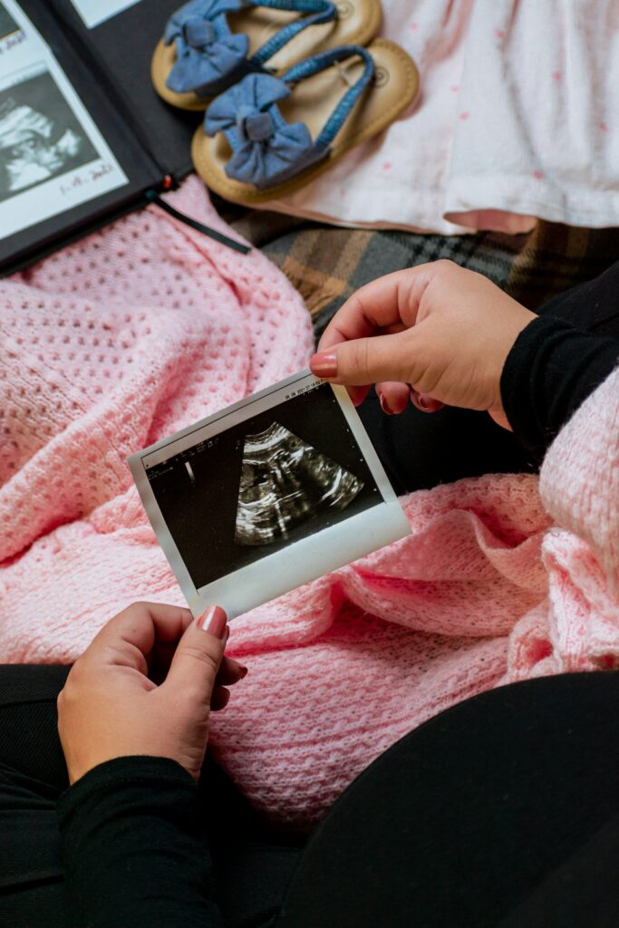
Ultrasound images depend on more than the equipment used. The baby’s position, gestational age, amount of amniotic fluid, maternal anatomy, and even the timing of the appointment can affect how clearly facial features, hands, feet, and other structures appear.
For families in Denison, TX, a few practical steps can improve the chances of obtaining useful images. These steps cannot guarantee a particular view, because fetal position and movement are not fully controllable, but they can help create favorable conditions.
What can be done before the ultrasound?
The most helpful preparation is following the instructions provided for the specific type of scan. Different examinations may require different bladder instructions, food restrictions, or medication guidance.
For many early-pregnancy abdominal ultrasounds, a partially full bladder may help provide a better viewing window. The bladder can lift the uterus slightly and improve the path of sound waves. Later in pregnancy, a full bladder is often unnecessary and may make the examination uncomfortable. Instructions should take priority over general advice.
Before leaving home, consider these simple steps:
- Wear comfortable clothing that allows easy access to the lower abdomen.
- Bring any requested records or information about pregnancy dates.
- Arrive with enough time to use the restroom or follow bladder instructions without rushing.
- Avoid applying thick oils, lotions, or creams to the abdomen immediately before the scan unless the imaging office specifically says they are acceptable.
- Eat and drink according to the instructions given for the appointment.
A heavy meal does not reliably make a baby move into a better position. Some people notice more fetal activity after eating or drinking something cold, but this varies widely and should not replace medical instructions.
Does the timing of the appointment affect image quality?
Yes. The stage of pregnancy strongly influences what can be seen. Early scans may show the pregnancy, gestational structures, and developing anatomy, while later scans may provide clearer views of the face, limbs, and body contours. However, a larger baby has less room to move, which can make certain views more difficult.
There is no universally perfect week for every image. The most useful timing depends on the purpose of the examination:
- An early diagnostic scan may focus on dating, location, growth, or viability.
- A first-trimester screening scan may require specific measurements during a limited developmental window.
- A detailed anatomy scan examines organs, limbs, growth, placenta, and other structures.
- A later scan may assess growth, position, fluid, or other pregnancy concerns.
If the goal is a particular keepsake view, it may be easier to obtain one during a period when the baby still has enough room to change position. Diagnostic examinations should always take priority over obtaining a certain facial image.
How does the baby’s position change the pictures?
Fetal position is one of the largest factors affecting ultrasound images. A baby facing the spine may be difficult to view from the front. Hands near the face, crossed legs, an arm across the chest, or the umbilical cord near the face can partially block important features.
The person performing the scan may ask the pregnant patient to change position, walk briefly, or wait for the baby to move. These adjustments can sometimes improve the angle, but movement is unpredictable. A baby may shift immediately, remain in the same position, or move away from a view that was previously clear.
A short break can occasionally help. The uterus may change slightly with movement, and fetal activity may increase naturally. Repeated repositioning is not always useful, especially if the examination is being performed for medical reasons or if the patient is uncomfortable.
Do food, cold drinks, or movement improve images?
They may influence fetal activity, but there is no dependable method that guarantees better images. A light snack, normal hydration, or a short walk may be suggested in some situations, but these approaches are not universally necessary.
Large amounts of sugar are not required and may be inappropriate for people who have been given dietary restrictions or who are monitoring blood glucose. Drinking excessive water can also create discomfort without improving the scan.
A practical approach is to follow the preparation instructions, remain reasonably comfortable, and allow time for natural fetal movement. If an image is difficult to obtain, the limitation is usually related to position, anatomy, gestational age, or the angle of the sound waves rather than something the patient did incorrectly.
Why might the images look unclear even when everything is normal?
Ultrasound does not work like a camera taking a direct photograph. It creates images from sound waves traveling through the body. Sound waves may be affected by tissue thickness, scar tissue, the position of the uterus, fetal movement, low or high amniotic fluid, and the distance between the probe and the area being examined.

Image clarity can also vary depending on whether the scan is performed through the abdomen or with an internal probe. An internal ultrasound may provide a closer view during early pregnancy, while an abdominal scan may be more appropriate as the pregnancy progresses.
An unclear picture does not automatically indicate a problem. It may simply mean that the baby is facing away, moving frequently, or positioned behind another structure. If a medically important area cannot be evaluated, additional imaging may be recommended for clinical reasons.
What should patients know about 3D and 4D images?
Three-dimensional ultrasound uses multiple two-dimensional images to create a surface rendering. Four-dimensional ultrasound adds movement over time. These formats can produce recognizable views of the face or limbs, but they are still affected by fetal position and the surrounding environment.
A clear 3D or 4D facial image generally requires:
- The face to be turned toward the probe.
- Enough amniotic fluid between the face and the uterine wall.
- The hands, feet, placenta, or umbilical cord not blocking the view.
- Limited movement during the portion of the scan used to create the rendering.
A baby’s face may appear flattened, shadowed, or distorted if it is pressed against the uterine wall. That appearance does not predict how the baby will look after birth. Facial images are also not a substitute for a diagnostic examination, and a pleasant-looking image cannot confirm that all anatomy is normal.
How can seasonal conditions affect comfort during a local appointment?
In Denison, hot and humid periods can make travel and waiting less comfortable, while colder weather may encourage people to wear layers that are difficult to adjust during an abdominal examination. Comfortable clothing, reasonable hydration, and planning extra travel time during severe weather can reduce stress before the scan.
The weather itself does not usually determine ultrasound quality. Comfort matters because remaining relaxed and still can make the examination easier, but patients should not delay medically necessary imaging because of ordinary seasonal conditions. During periods of flooding, storms, or dangerous roads, appointment instructions and personal safety should guide travel decisions.
What should happen if the baby will not cooperate?
A baby who does not move into the desired position is common. The imaging professional may try a different angle, ask for a position change, wait briefly, or explain that a particular view cannot be captured at that time.
If the scan is diagnostic, the important question is whether the required anatomy and measurements were adequately evaluated. If a specific medical view remains incomplete, follow-up imaging may be arranged. If the scan is primarily intended for optional keepsake images, the result may simply be limited by fetal position.
Patients can ask which images were successfully obtained, whether any medically necessary views were incomplete, and whether additional imaging is recommended. Those questions provide more useful information than judging the scan solely by the appearance of a single printed or digital picture.
Which expectations are realistic?
The clearest images usually come from realistic expectations, proper preparation, and patience with factors that cannot be controlled. Not every appointment produces a profile, open eyes, visible fingers, or a full facial view.
The central purpose of prenatal imaging is assessing pregnancy development and maternal-fetal health. The most memorable images may be the ones that happen naturally, but a less dramatic picture can still document an important moment in the pregnancy and provide valuable clinical information.