

 See My Latest Article on HelloNation
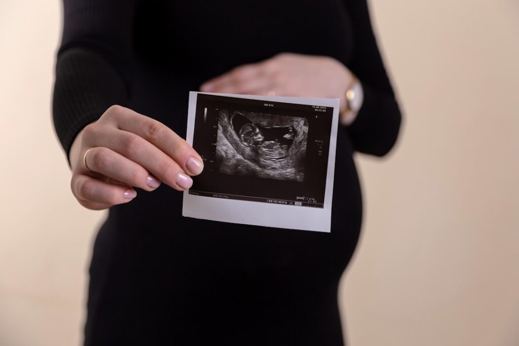
What Is Prenatal Imaging and What Purposes Does It Serve?
Prenatal imaging refers to medical scans that allow healthcare providers to view and assess a developing baby and the health of the pregnancy. The most recognized form is ultrasound, though some pregnancies may require additional scans or imaging techniques depending on specific circumstances. Local clinics in Denison typically use non-invasive technologies, making the process safe for both mother and baby in most situations.
Prenatal imaging provides a window into early development, helping families and healthcare teams answer key questions about a pregnancy’s progress and the well-being of both mother and child.
How Does Prenatal Imaging Help Mothers in Denison?
Prenatal imaging helps mothers by offering reassurance, guiding decision-making, and improving overall pregnancy care. In the Denison climate, where families often balance work, outdoor activities, and seasonal weather shifts, knowing what’s happening in a pregnancy offers peace of mind and supports healthy planning.
Two key ways local mothers benefit include:
- Early confirmation of a viable pregnancy.
- Monitoring for common issues such as gestational age, multiple pregnancies (twins or more), or abnormalities in the uterus.
Carefully-timed scans can also help identify conditions like placenta previa or low amniotic fluid, which are easier to address when caught early. For residents who might face longer travel times to major hospitals, timely information empowers families to prepare and seek further care if needed.
How Does Prenatal Imaging Support Baby’s Health?
Prenatal imaging allows providers to monitor the baby's growth, assess organ development, and spot issues before birth. For example, measuring the baby’s size and comparing it to expected milestones helps signal whether the baby is thriving. Identifying developmental or structural concerns—such as heart defects or spinal issues—gives medical teams a valuable head start to coordinate specialized care.
In rural or semi-rural areas like Denison, advanced notice of potential complications is key, since access to specialized care may require additional coordination or travel. Early detection doesn’t just improve outcomes—it allows time for families to learn and plan for any extra support their baby might need.
What Are the Main Types of Prenatal Imaging Used Locally?
Most pregnant residents in Denison will have at least one standard ultrasound (sometimes called a “sonogram”) during their pregnancy. Ultrasounds use sound waves to create real-time images of the baby and the pregnancy environment. Common types include:
- First-trimester ultrasound: Typically done between 6-12 weeks, this scan confirms pregnancy, estimates due date, and checks the baby’s heartbeat.
- Second-trimester “anatomy scan": Usually between 18-22 weeks, this detailed scan checks organ development, assesses placenta placement, and can reveal the baby’s sex.
- Additional ultrasounds: Used if concerns arise or to monitor specific health issues during later trimesters.
Other imaging, such as specialized Doppler studies or MRI, is far less common but may be suggested by a healthcare provider for complex cases.
How Often Should Pregnant Women Expect These Tests?
In standard pregnancies, two or three ultrasounds are routine. Timing and frequency depend on health conditions, medical history, or any risk factors present. For example, women with diabetes, high blood pressure, or previous pregnancy complications may require more frequent scans.
Communication with local healthcare providers ensures scans are scheduled appropriately, reflecting individual health and the specific needs of the pregnancy.
Are Prenatal Imaging Procedures Safe?
Ultrasound, the primary form of prenatal imaging, is considered safe for both mother and baby when performed by trained staff following medical guidelines. Unlike X-rays, ultrasound does not use ionizing radiation, making it preferable for ongoing monitoring.

Misconceptions sometimes arise about the risks of repeated imaging. However, routine ultrasounds, as used in Denison and throughout the U.S., do not harm the developing baby or the mother. That said, imaging should always be guided by a medical need and performed by qualified professionals.
What Information Is Provided by These Scans?
Prenatal scans can clarify:
- The age and expected due date of the baby
- Number of babies present
- Major physical developments, including heart, brain, limbs, and spine
- Location of the placenta and amount of amniotic fluid
- Clues about genetic or developmental conditions (though not all disorders are visible)
This information helps parents anticipate needs, make informed choices, and work with providers to plan for birth—whether delivery is anticipated at home, in a hospital, or at a specialized center outside Denison if necessary.
What Are the Limitations of Prenatal Imaging?
While prenatal imaging is highly useful, no scan can guarantee a baby will be completely healthy, nor can every condition be detected before birth. Some issues develop later or may be too subtle to appear on an image. Anatomical scans may suggest—but cannot confirm—the presence of genetic conditions. Imaging should be part of a broader conversation about prenatal health.
Local weather or power interruptions can sometimes cause appointment delays, so families in the area may want to plan for flexibility, especially during severe spring weather or ice storms.
How Can Families Use Prenatal Imaging Results?
Scan results should be discussed with a provider who knows the family's history and situation. For some, images offer simple reassurance to continue with routine prenatal care. For others, results might prompt additional testing, medication adjustments, or birth planning. Some families use these findings to involve specialists or access local support resources if potential health issues are identified early.
Understanding what imaging can—and cannot—provide helps set realistic expectations and guides meaningful preparation for a healthy pregnancy journey in Denison.