

 See My Latest Article on HelloNation
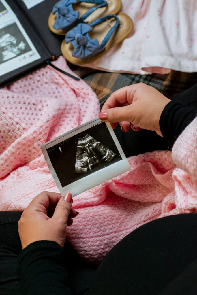
What Is Prenatal Imaging and Why Is It Used?
Prenatal imaging refers to a range of medical techniques used to examine and monitor an unborn baby during pregnancy. The main purpose is to help both healthcare teams and expectant families track development, detect certain conditions, and provide information about the pregnancy’s progress. For residents of Denison, TX, these imaging methods follow national guidelines but may be tailored to the rhythms and resources of local healthcare, making knowledge of each type especially valuable.
What Are the Main Types of Prenatal Imaging?
The most familiar prenatal imaging method is ultrasound, but several others exist for specific situations.
Ultrasound Imaging
Ultrasound is the routine cornerstone of imaging during pregnancy. It uses safe, high-frequency sound waves to create live images of the baby, placenta, amniotic fluid, and uterus.
- Standard (2D) Ultrasound: Most pregnancies receive at least one standard ultrasound, typically around 18-22 weeks. This exam provides a comprehensive check of the baby's anatomy and the pregnancy environment.
- Early Ultrasound: Performed in the first trimester, often to confirm pregnancy, estimate a due date, and check for multiples.
- Advanced (3D or 4D) Ultrasound: Creates more detailed pictures or moving images. While sometimes used to clarify findings, these are not a routine necessity.
Doppler Ultrasound
Different from standard ultrasound, Doppler ultrasound measures the movement of blood through the baby's heart or blood vessels. In Denison, this type may be used if there are concerns about fetal growth or circulation, such as in pregnancies affected by high blood pressure or suspected fetal anemia.
Specialized Imaging: When Is It Used?
Other imaging types are reserved for specific medical indications and are not generally part of routine pregnancy care.
- Fetal Echocardiography: A targeted scan of the baby's heart, often recommended if a previous child has a heart condition, or if a possible abnormality is detected during a regular ultrasound.
- Magnetic Resonance Imaging (MRI): Very rarely needed, but occasionally used in complex cases to get highly detailed images of organs or structures if ultrasound does not provide enough detail.
How Are These Imaging Studies Scheduled in Denison?
Local healthcare providers coordinate prenatal imaging based on medical guidelines, available equipment, and each patient’s unique circumstances. Most people in the area have their main ultrasound performed during a second trimester prenatal appointment. Additional or specialized imaging is arranged if there are medical questions that cannot be answered through routine exams.
Are Prenatal Imaging Methods Safe for My Baby and Me?
For families in Denison, safety is a frequent concern. Ultrasound, including standard and Doppler types, is considered very safe because it does not use radiation. MRI is also safe in pregnancy if required, as it uses strong magnets and radio waves instead of X-rays.
Residents sometimes worry about possible risks of unnecessary or repeated exams. Local practitioners aim to balance medical need with minimizing any unhelpful tests; non-essential imaging such as keepsake 3D/4D scans is generally not recommended unless there’s a clinical reason.
What Will Happen During My Imaging Appointment?
Prenatal imaging appointments are designed to be as comfortable as possible. A typical ultrasound takes about 20-30 minutes. Expect:
- A cool gel will be applied to the abdomen
- A handheld device called a transducer glides over the skin, sending images to a monitor
- Occasionally, a transvaginal approach is used very early in pregnancy for better images
Actual procedures may vary slightly depending on the stage of pregnancy and the questions being answered.

Can Imaging Screens Detect All Potential Concerns?
Prenatal imaging is a powerful tool, but it has limits. Not all conditions can be seen through these methods. Local providers in the city use imaging to:
- Detect certain structural problems
- Estimate growth and development
- Monitor certain health risks
However, some conditions and subtle fetal changes may not be visible even on the most advanced scans. False positives (worrisome findings that turn out to be normal) can occasionally occur, causing temporary anxiety for families.
Are There Special Local Considerations in Denison?
Denison’s population includes both suburban households and those from rural surroundings, impacting imaging access. Not all area clinics offer every type of prenatal imaging, especially more advanced tests such as MRI or fetal echocardiography. In these cases, some families may travel to larger hospitals outside the immediate area for certain referrals.
Severe weather and seasonal storms can occasionally disrupt travel to imaging appointments. During winter storms or periods of high rain, local clinics may adjust their schedules for safety, and area residents are encouraged to plan visits with these factors in mind.
Common Misconceptions About Prenatal Imaging
It’s a myth that prenatal imaging guarantees “perfect” health or predicts every possible issue. Some believe that more imaging equals better care, but medical guidelines recommend tests only when there’s a valid reason. In Denison, asking questions during appointments and sharing family history can help ensure the right types of imaging are chosen.
When Might Imaging Be Repeated or Increased?
Certain pregnancies need closer follow-up, which may include additional imaging. Examples include:
- Carrying multiples (such as twins)
- Having medical conditions like diabetes or hypertension
- Previous pregnancy issues, such as preterm birth or birth defects
In such cases, additional ultrasounds or specialized imaging may be planned to provide the best information for the health of both mother and baby.