

 See My Latest Article on HelloNation
How Does Prenatal Imaging Help Identify Birth Defects?
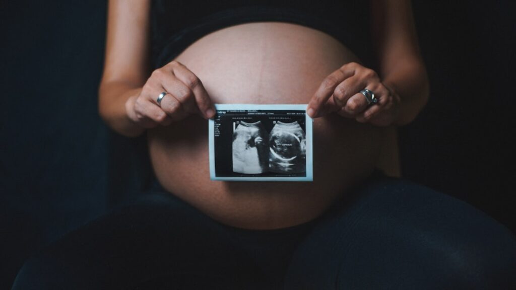
Prenatal imaging provides a safe, noninvasive window into a baby’s development during pregnancy and is the primary method for early detection of many structural birth defects. By visualizing a baby’s organs, limbs, and overall growth, doctors can identify concerns months before birth, allowing families and healthcare teams to better prepare for the baby’s arrival.
These imaging tests are standard for most pregnancies, offering reassurance about typical development while also flagging any variations that may need attention.
Which Tests Are Commonly Used in Pregnancy?
The main prenatal imaging tool is the ultrasound. During an ultrasound, sound waves create moving pictures of the fetus inside the womb. Most expectant parents in the city have at least two ultrasounds: one in the first trimester to confirm the pregnancy and estimate due dates, and a second, detailed scan around 18-22 weeks to carefully examine the baby's anatomy.
Other imaging tests sometimes used include:
- Blood tests combined with ultrasound for increased accuracy when screening for certain chromosomal abnormalities
- Fetal echocardiography for close examination of the baby’s heart structure and function, especially if there’s an increased risk of heart defects
MRI is occasionally recommended if an ultrasound shows something unusual and more information is needed, though it is less common during pregnancy.
What Birth Defects Can Be Detected with Prenatal Imaging?
Many—but not all—birth defects can be identified with imaging. Common conditions detected include:
- Neural tube defects (like spina bifida)
- Abdominal wall defects
- Heart defects
- Kidney and urinary tract abnormalities
- Cleft lip and palate
- Limb and bone abnormalities
Some conditions, such as milder heart defects or certain genetic syndromes, may not be visible on imaging alone. Also, certain problems may develop later in pregnancy or may only become apparent after birth.
Why Is Early Detection Important for Area Families?
Knowing about a birth defect before delivery gives families time to learn about the condition, connect with resources, and plan for specialized medical needs. In certain cases, early detection affects delivery planning or enables immediate care that improves outcomes for the baby.
For community members in Denison with access to both rural and suburban amenities, early information can also help arrange necessary travel or coordinate care at larger facilities if specialized support will be needed at birth.
Are Prenatal Imaging Tests Safe?
Ultrasound is considered very safe for pregnant individuals and their babies. No use of ionizing radiation occurs during ultrasound, and it has been used routinely for decades. MRI is also used safely in pregnancy but is generally reserved for very specific indications.
Families sometimes worry about the possibility of overexposure or unnecessary scans. In local practices, ultrasounds are only ordered when medically needed, and providers avoid unneeded imaging.
What Should Expectant Parents Know About the Process?
Prenatal ultrasounds in Denison are typically performed in outpatient clinics, medical centers, or hospital-based settings. Routine scans are painless, take about 20-45 minutes, and may involve a full bladder for the clearest images early in pregnancy.
Misconceptions sometimes circulate about what ultrasounds can and cannot find. Ultrasounds are excellent for structural information—seeing organs, limbs, and growth—but cannot diagnose all genetic or metabolic conditions. No test guarantees a completely “normal” baby, though most families find reassurance from normal imaging results.

What Happens If a Concern Is Found?
If a prenatal imaging scan finds something unusual, further steps may include:
- Additional ultrasounds to monitor changes over time
- Referral to a maternal-fetal medicine specialist for detailed assessment
- Meetings with pediatric specialists or genetic counselors to discuss possible causes and outcomes
In some cases, expectant parents may be offered additional testing, such as amniocentesis, to clarify the diagnosis. Emotional support and clear communication are also critical. Many families in the area benefit from local and regional support resources if facing a new health challenge.
Can Anything Be Missed by Prenatal Imaging?
No test is perfect. Some birth defects are subtle or develop later in pregnancy and may not be picked up during routine scans. In pregnancies with factors such as higher body mass index, limited fetal movement, or certain anatomical positions, images may not be as clear. Providers in Denison always discuss the limitations honestly so families know what to expect.
What Questions Should Residents Ask Their Healthcare Providers?
To get the most out of the prenatal imaging experience, local residents may want to ask:
- Which routine scans are recommended for my pregnancy?
- What kinds of birth defects can and cannot be detected?
- If something unusual is found, what steps are next?
- Are there support resources available if a health concern arises?
- Who will explain the results in detail?
By understanding these aspects, families in the community are empowered to participate actively in their prenatal care and remain informed about their options.